Division of Biology and Medicine
Center for Alternatives to Animals in Testing
Core Resources
We use quantitative confocal imaging of 3D human microtissues to identify pathologic responses to chemical and drug exposures.
Core Resources
We use quantitative confocal imaging of 3D human microtissues to identify pathologic responses to chemical and drug exposures.
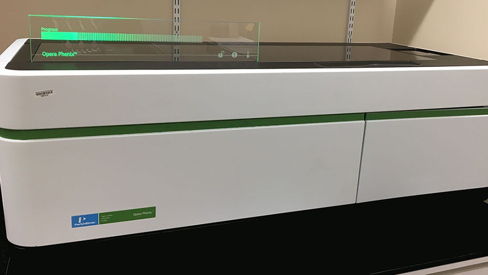
Opera Phenix High-Content Screening System
The Opera Phenix High-Content Screening System is a microscope designed for high-throughput 3D imaging of fluorescent samples in microplates. This imaging system, open to all investigators, is critical to the mission of the Center for Animal Alternatives in Testing and associated NSF/EPSCoR and NIH/BRP awards.
1
of
1