Breast
The Boekelheide lab is using breast 3D microtissues to investigate the toxicological effects of estrogens and chemicals that mimic estrogen’s effects.
Breast
The Boekelheide lab is using breast 3D microtissues to investigate the toxicological effects of estrogens and chemicals that mimic estrogen’s effects.
Breast 3D Microtissues
- The Boekelheide lab has a developed a 3D microtissue of the human breast. The microtissue is comprised of human breast cancer cells.
- These cells aggregate to form a single 3D microtissue that then undergoes an additional and dramatic morphological change. The single initial microtissue forms several smaller clusters of cells, each with a lumen or hollow space. Each lumen is surrounded by a cell layer 1-2 cells thick.
- We used a periodic acid-Schiff stain and showed that carbohydrate-positive material is being secreted into the lumens. Electron microscopy revealed the presence of microvilli, small finger-like cellular projections on the interior surface of the lumens.
- This demonstrates that in addition to morphogenesis, the cells are undergoing polarization, with their apical surface facing the luminal space and their basal-like surface facing the exterior of the microtissue.
- We also showed that the breast microtissues have strong staining for mucin 1 and milk fat globule-EGF factor 8 protein and strong staining for E-cadherin localized to the cell-cell contacts.
- A comparison of gene and protein expression between cells in a 2D monolayer versus the same cells in a 3D microtissue showed that the 3D environment significantly enhanced the expression of genes associated with differentiation and more closely mimicked that of breast tissue.
- To determine if the breast microtissues were responsive to hormone, we treated the microtissues with 17β-estradiol for 8 hours, and analyzed the expression of genes known to be targets of estrogen. Treatment with 17β-estradiol caused a dose-dependent increase in expression of the GREB gene (growth regulation by estrogen in breast cancer 1) and the PGR gene (progesterone receptor).
- We are using human breast 3D microtissues to investigate the toxicological effects of estrogens and chemicals that mimic estrogenic’s effects.
 Play
Play
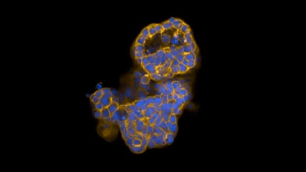
3D morphology of breast microtissue stained with nuclear stain (blue) and actin stain (yellow).
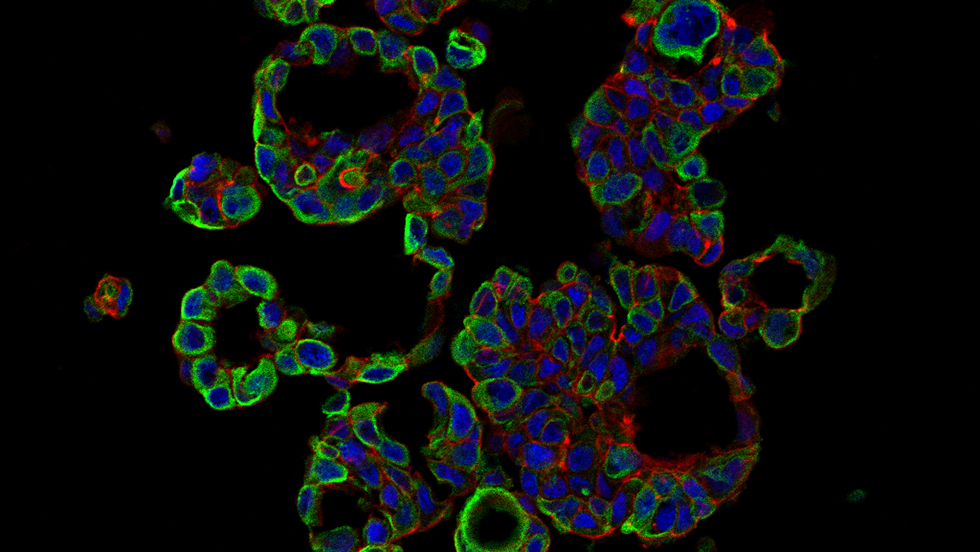
MCF-7 microtissues grown for 7 days exhibit expression of keratin 8 (green), a luminal epithelial marker. Counterstained with DAPI (blue) and rhodamine phalloidin (red).
Learn More
Vantangoli, M. M., Wilson, S., Madnick, S. J., Huse, S. M., & Boekelheide, K. (2016). Morphologic effects of estrogen stimulation on 3D MCF-7 microtissues. Toxicology Letters, 248, 1-8. PMC4803074
Vantangoli, M. M., Madnick, S. J., Wilson, S., & Boekelheide, K. (2016). Estradiol exposure differentially alters monolayer versus microtissue MCF-7 human breast carcinoma cultures. PLoS One, 11(7), e0157997. PMC4933361
Vantangoli, M. M., Madnick, S. J., Huse, S. M., Weston, P., & Boekelheide, K. (2015). MCF-7 Human breast cancer cells form differentiated microtissues in scaffold-free hydrogels. PLoS One, 10(8), e0135426. PMC4534042
Investigators
-

Kim Boekelheide, MD, PhD
Associate Director of the Center for Animal Alternatives in Testing, Professor of Medical Science, Department of Pathology and Laboratory Medicine