Ovary
The Morgan lab is using ovary microtissues to measure the effects of drugs and environmental chemicals that can affect fertility.
Ovary
The Morgan lab is using ovary microtissues to measure the effects of drugs and environmental chemicals that can affect fertility.
Ovary 3D Microtissues
- The Morgan lab has developed a 3D microtissue of the human ovary. The microtissue is comprised of human granulosa cells, the cells that surround and nourish the oocyte (immature egg) in the female ovary.
- The ovary microtissue is a multi-cell layered structure that mimics the avascular architecture of the granulosa surrounding the oocyte.
- The cells in the microtissue form functional gap junctions, direct connections that electrically and chemically couple the cells. Gap junction intercellular communication relays important signals for the maturation of the oocyte.
- We have utilized the Opera Phenix high-content imaging system to develop a high throughput assay to measure gap junction intercellular communication in living 3D microtissues.
- The ovary microtissues also synthesize and secrete estradiol and progesterone, two hormones that are critical for oocyte maturation and successful reproduction.
- Estradiol synthesis by the microtissue is induced by follicle stimulating hormone and cAMP and we’ve shown that it is inhibited by chemicals that target the aromatase enzyme (CYP19) central to the synthesis of estradiol.
- We are using ovary microtissues to measure the effects of drugs and environmental chemicals that can affect fertility by disrupting gap junction communication and the synthesis of estradiol.
Three-dimensional rendering of a human ovary microtissue reconstructed from a z-stack of images from the confocal microscope. Only 10% of the cells were labeled with a fluorescent dye so this video shows the size and shape of individual cells within a 3D microtissue.

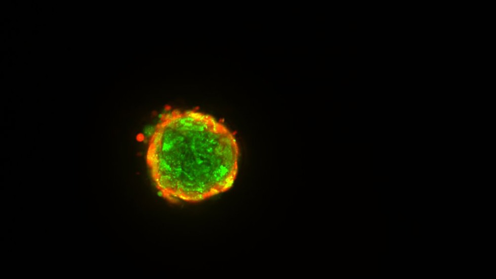
A living ovary microtissue stained with two fluorescent dyes for the measurement of gap junction communication between cells. All cells are stained with the green dye. The red dye is absorbed by the microtissue and passes into its center via the small pores of gap junctions that connect cells.
Learn More
Bao, B., Jiang, J., Yanase, T., Nishi, Y., & Morgan, J. R. (2011). Connexon-mediated cell adhesion drives microtissue self-assembly. FASEB Journal, 25(1), 255-264. PMC3005422
Leary, E., Rhee, C., Wilks, B., & Morgan, J. R. (2016). Quantitative wide field fluorescence microscopy of 3D spheroids. BioTechniques, 61(5), 237-247. PMID: 27839509
Leary, E., Rhee, C., Wilks, B. T., & Morgan, J. R. (2018). Quantitative live-cell confocal imaging of 3D spheroids in a high-throughput format. SLAS Technology, 23(3), 231-242. PMC5962438
Investigators
-

Jeffrey Morgan, PhD
Donna Weiss ’89 and Jason Weiss Director of the Center for Alternatives to Animals in Testing, Professor of Medical Science, Department of Pathology and Laboratory Medicine -

Blanche Ip, PhD
Assistant Professor of Pathology and Laboratory Medicine