3D Cell Culture Environments
3D microtissues mimic the function, morphology and cell density of natural tissues and organs and so provide a platform for the development of more predictive assays for drug discovery and toxicity testing.
3D Cell Culture Environments
3D microtissues mimic the function, morphology and cell density of natural tissues and organs and so provide a platform for the development of more predictive assays for drug discovery and toxicity testing.
3D Cell Culture Technology
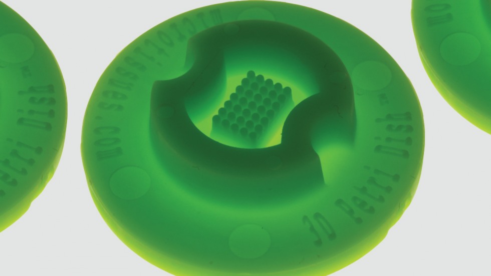
- The Morgan lab invented the 3D PetriDish®, now commercially available (Sigma-Aldrich), to grow cells in 3D using micro-molded agarose gels.
- Unlike other methods in tissue engineering that attach cells to a scaffold, cells are seeded into micro-molded nonadhesive agarose gels where they self-assemble an array of 3D multi-cellular microtissues in typically 24 hours.
- The platform technology works for a wide variety of cell types from cell lines to primary cells from numerous tissues and organs including those from human.
- The transparent micro-molded agarose provides a stable environment for long term (weeks) culture that is suitable for quantitative microscopy.
- These 3D microtissues mimic the function, morphology and cell density of natural tissues and organs and so provide a platform for the development of more predictive assays for drug discovery and toxicity testing.
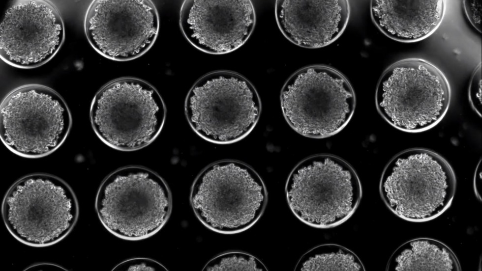
- Many of the investigators in the Center use this platform technology to make microtissues mimicking various organ systems.
- The Morgan lab is investigating several areas including an ovary 3D microtissue for toxicity testing, the function of gap junctions, the role of drug efflux pumps and the development of quantitative confocal image-based high throughput 3D assays.
Many researchers at the Center use the 3D Petri Dish® as a platform to construct microtissues that mimic various organ systems.
 Play
Play
Time lapse video of human stem cells aggregating and self-assembling into 3D microtissues, one per micro-well. Elapsed time of video is twenty hours.
Learn More
Napolitano, A.P., Chai, P., Dean, D.M., Morgan, J.R. (2007). Dynamics of the self-assembly of complex cellular aggregates on micro-molded non-adhesive hydrogels. Tissue Engineering, 13(8), 2087-2094. PMID: 17518713
Napolitano, A.P., Dean, D.M., Man, A.J., Youssef, J., Ho D.N., Rago, A.P, Lech, M.P., Morgan, J.R. (2007). Scaffold-free 3-dimensional cell culture utilizing micro-molded non-adhesive hydrogels. BioTechniques, 43, 494-500. PMID: 18019341
Barbone, D., Yang, T-M, Morgan, J.R., Gaudino, G., Broaddus, V.C. (2008). mTOR contributes to the acquired multicellular apoptotic resistance of human malignant mesothelioma spheroids. Journal of Biological Chemistry. 283(19), 13021-13030. PMC2442321
Bao, B., Jiang, J. Yanase, T., Nishi, Y., and Morgan, J.R. (2011) Connexon-mediated cell adhesion drives microtissue self-assembly. FASEB Journal, (25)1, 255-264. PMC3005422
Achilli, T-M., McCalla, S., Tripathi, A. and Morgan, J.R. (2012). Quantification of the kinetics and extent of self-sorting in three dimensional spheroids. Tissue Engineering, 18(4), 302-309. PMC3312373
Bao, B.A., Lai, C.P.K., Naus, C.C., and Morgan, J.R. (2012). Pannexin 1 drives multicellular compaction via a signaling cascade that upregulates cytoskeletal function. Journal of Biological Chemistry, 287(11), 8407-8416. PMC3318751
Desroches, B.R., Zhang, P., Choi, B., Maldonado, A.E., Rago, A., Liu, G.X. Nath, N., King, M.E., Hartmann, K.M., Yang, B., Koren, G., Morgan, J.R. and Mende U. (2012). Functional scaffold-free cardiac microtissues: A novel model for the investigation of heart cells. American Journal of Physiology Heart and Circulatory Physiology, 302(10), H22031-H220442. PMC3362102
Achilli, T-M, McCalla, S., Meyer, J., Tripathi, A., Morgan, J.R. (2014). Multilayer spheroids to quantify drug uptake and diffusion in 3D. Molecular Pharmaceutics 11(7), 2071-2081. PMC4096226
Curran, S., Achilli, T-M, Leary, E., Wilks, B., Vantangoli, M.M., Boekelheide, K., and Morgan, J.R. (2015). A 3D spheroid system to evaluate inhibitors of the ABCG2 transporter in drug uptake and penetration. Technology, 3, 54-63, 2015. doi:10.1142/S2339547815500028
Curran, S., Vantangoli, M. M., Boekelheide, K., and Morgan, J.R. (2015). Architecture of chimeric spheroids controls drug transport. Cancer Microenvironment, 8(2), 101-109, 2015. PMC4542826
Kabadi, P.K., Vantangoli, M.M., Rodd, A.L., Leary, E., Madnick, S.J., Morgan, J.R., Kane, A. and Boekelheide, K. (2015). Into the depths: Techniques for in vitro three-dimensional microtissue visualization. BioTechniques, 59(5), 279-86. PMC4804457
Dingle, Y.T., Boutin, M.E., Chirila, A.M., Livi, L.L., Labriola, N.R., Jakubek, L.M., Morgan, J.R., Darling, E.M., Kauer, J.A., Hoffman-Kim, D. (2015). Three-dimensional neural spheroid culture: an in vitro model for cortical studies. Tissue Engineering Part C Methods, 21(12),1274-83. PMC4663656
Leary, E., Rhee, C., Wilks, B., and Morgan, J.R. (2016). Quantitative wide field fluorescence microscopy of 3D spheroids. BioTechniques, 61, 237-247, 2016. PMID: 27839509
Leary, E., Rhee, C., Wilks, B. T., and Morgan, J.R. (2018). Quantitative live-cell confocal imaging of 3D spheroids in a high-throughput format. SLAS Technology, 23(3), 231-242. PMC5962438.
Investigators
-
Jeffrey Morgan, PhD
Donna Weiss ’89 and Jason Weiss Director of the Center for Alternatives to Animals in Testing, Professor of Medical Science, Department of Pathology and Laboratory Medicine