Optical Coherence Tomography: Label-free Measurements of Microtissue Viability
Researchers led by Professor Jonghwan Lee are developing 3D microscopic imaging and image processing technologies for label-free assessment of the viability of cells in 3D microtissues.
Optical Coherence Tomography: Label-free Measurements of Microtissue Viability
Researchers led by Professor Jonghwan Lee are developing 3D microscopic imaging and image processing technologies for label-free assessment of the viability of cells in 3D microtissues.
Optical Coherence Tomography
- Optical coherence tomography (OCT) is used routinely in the clinic to create 3D images of the human retina and diagnose disease. The Lee Lab is using OCT to image 3D human microtissues.
- OCT imaging harnesses the properties of light to create quantitative 3D images of microtissues. Unlike other imaging modalities, OCT imaging is nondestructive. There is no need to add fluorescent dyes or labels and the same microtissues can be repeatedly imaged over weeks.
- The Lee Lab has advanced the OCT technology beyond imaging structures and is now able to do quantitative imaging and assessment of multiple dynamic processes that occur in living microtissues.
- One such advance is the combination of OCT with dynamic light scattering, called DLS-OCT, which produces a 3D map of the movements inside living cells. Since movement inside cells is governed by the metabolism of the cell, DLS-OCT images can be used to indirectly assess the viability of cells.
- In addition to metabolic activity, the Lee Lab is evaluating a comprehensive set of image features to assess how drugs and toxicants disrupt specific cellular pathways including integrity of the cell membrane and the cytoskeleton.
- The goal of the Lee Lab is to develop OCT imaging as a rapid imaging tool for the high-throughput analysis of the viability of human microtissues, thus improving the ease, cost and speed of the safety testing of chemicals as well as the testing of the efficacy and safety of new drugs.
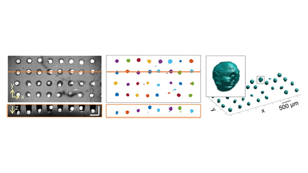
Top and side view of OCT images of an array of brain microtissues (left) and automated segmentation and highlighting of the brain microtissues (center) and 3D-rendered view of the brain microtissues (right).
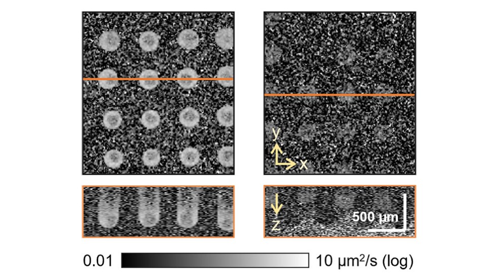
DLS-OCT imaging of live brain microtissues (left) and dead microtissues killed by fixation (right). The live microtissues exhibit higher diffusion coefficients that reflect the intracellular motility which is governed by the level of cellular metabolic viability.
Learn More
J Lee, W Wu, JY Jiang, B Zhu, DA Boas, “Dynamic light scattering optical coherence tomography”, Optics Express 20 (20), 22262-22277 (2012).
J Lee, H Radhakrishnan, W Wu, A Daneshmand, M Climov, C Ayata, DA Boas, “Quantitative imaging of cerebral blood flow velocity and intracellular motility using dynamic light scattering–optical coherence tomography”, Journal of Cerebral Blood Flow & Metabolism 33 (6), 819-825 (2013).
JS Lee, K Eom, C Polucha, J Lee, “Standard-unit measurement of cellular viability using dynamic light scattering optical coherence microscopy”, Biomedical Optics Express 9 (11), 5227-5239 (2018).
S Stefan, KS Jeong, C Polucha, N Tapinos, SA Toms, J Lee, “Determination of confocal profile and curved focal plane for OCT mapping of the attenuation coefficient”, Biomedical Optics Express 9 (10), 5084-5099 (2018).
Investigators
-

Jonghwan Lee, PhD
Assistant Professor of Engineering, School of Engineering, Center for Biomedical Engineering, Carney Institute for Brain Science