Lung
Lung microtissues are being developed as a tiered testing strategy and initial step for screening for the potential hazards of new nanomaterials.
Lung
Lung microtissues are being developed as a tiered testing strategy and initial step for screening for the potential hazards of new nanomaterials.
Human Lung 3D Microtissues
- The Kane lab in collaboration with the Hurt lab has developed a 3D microtissue of the human ling. The lung 3D microtissue is comprised of a mixture of human lung epithelial cells, human lung fibroblasts and human macrophages.
- The cells aggregate and form a stable lung 3D microtissue with epithelial cells and fibroblasts interspersed and macrophages located at the periphery.
- This mix of cells and 3D morphology represents the epithelial-mesenchymal trophic unit present in the lung during persistent inflammation, injury, aberrant repair and fibrosis. It also recapitulates the cellular cross talk and signaling that occurs between different cell types.
- We have treated the lung microtissues with asbestos, a well-known fiber that induces lung pathology, and compared that response to lung microtissues treated with multi-walled carbon nanotubes, a fiber that is a new and emerging engineered nanomaterial finding applications in the electronics industry. Both fibers were taken up by the lung microtissues and induced the expression of genes associated with persistent inflammation and fibrosis.
- These studies are providing mechanistic insights into the specific properties of engineered nanomaterials that may be responsible for adverse human health effects.
- We are developing lung microtissues as a tiered testing strategy and initial step for screening for the potential hazards of new nanomaterials.
 Play
Play
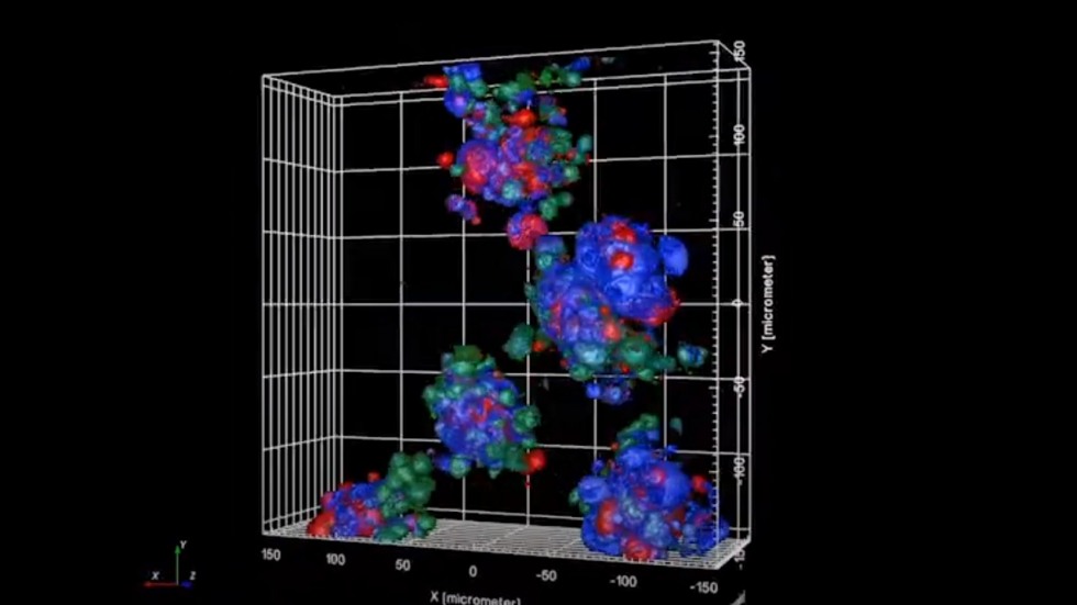
Triculture cell-cell interactions and organization was evaluated in live spheroids during and after formation. 3D microtissue cell types were stained with cytoplasmic (red, green, blue) and nuclear stains (white) and allowed to spontaneously form microtissues.
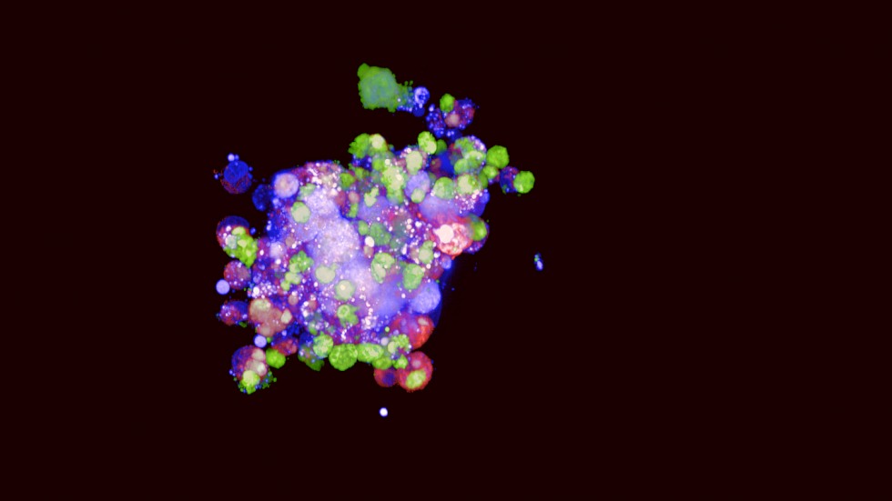
Fluorescent image of a human lung microtissue comprised of three cell types: lung bronchial epithelial cells (red), macrophages (green) and lung fibroblasts (blue).
Learn More
Kabadi, P.K., Rodd, A.L., Simmons, A.E., Messier, N.J., Hurt, R.H., Kane, A.B. (2019). A novel human 3D lung microtissue model for nanoparticle-induced cell-matrix alterations. Particle and Fibre Toxicology, 16(1), 15. PMC6448215
Kabadi, P. K., Vantangoli, M. M., Rodd, A. L., Leary, E., Madnick, S. J., Morgan, J. R., Kane, A., & Boekelheide, K. (2015). Into the depths: Techniques for in vitro three-dimensional microtissue visualization. Biotechniques, 59(5), 279-86. PMC4804457
Investigators
-

Agnes Kane, MD, PhD
Professor of Medical Science, Department of Pathology and Laboratory Medicine -

Robert Hurt, PhD
Professor of Engineering, School of Engineering