Fish Liver
Researchers are using 3D microtissues to study the effects of pollutants and engineered nanomaterials on the liver.
Fish Liver
Researchers are using 3D microtissues to study the effects of pollutants and engineered nanomaterials on the liver.
Fish Liver 3D Microtissues
- The Kane lab, in collaboration with the Hurt lab, has developed a 3D microtissue of fish liver. The microtissue is comprised of PLHC-1 liver cells from the fish, Poeciliopsis lucida.
- Over the course of three days, these cells self-assembled and aggregated into a compact homogenous microtissue wherein the cells adopted a cuboidal shape in contrast to their flattened appearance when grown as two-dimensional monolayers.
- Like normal liver, the fish liver microtissues accumulated intracellular glycogen granules and expressed the xenobiotic metabolizing enzyme cytochrome P450 1A (Cyp1a).
- When exposed to low doses and repeated doses of the model environmental toxicant benzo(a)pyrene, the fish liver microtissues underwent significant morphological changes and altered expression of CYP1a.
- We are using fish liver microtissues as an aquatic toxicity testing platform to investigate the effects of pollutants such as the polycyclic aromatic hydrocarbons found in petroleum as well as the use of new nanomaterials that are being engineered as alternatives to chemical-based dispersants used to remediate marine oil spills.
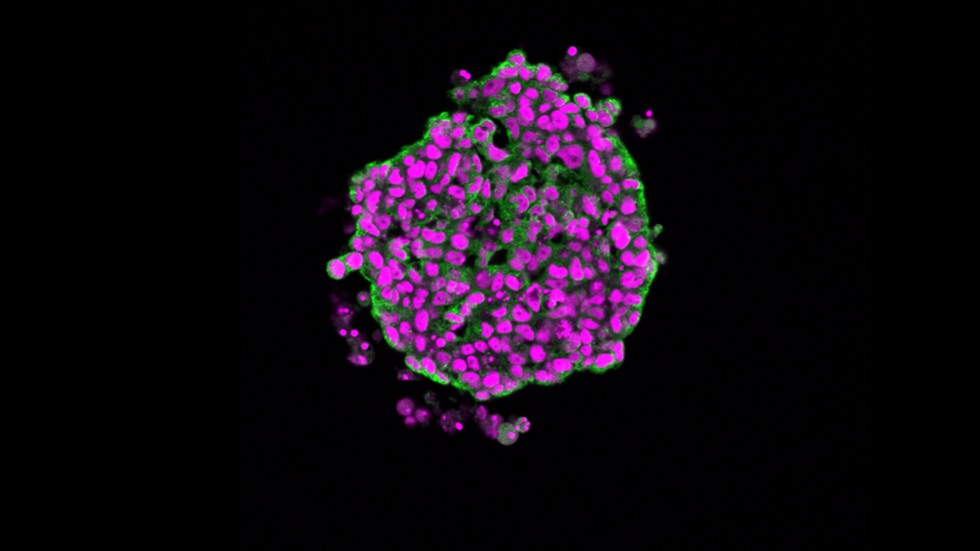
This fish liver microtissues can be used to measure biomarkers of exposure to pollutants like benzo(a)pyrene, including changes in the health of cells through morphological changes in the nuclei (magenta).
 Play
Play
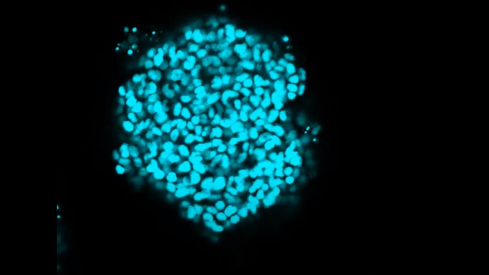
Video showing the nuclei of a 3D fish liver microtissue stained with Hoechst 3342 (blue). Video progresses through a stack of confocal images, each z-slice is 1 micron.
Learn More
Rodd, A. L., Messier, N. J., Vaslet, C. A., & Kane, A. B. (2017). A 3D fish liver model for aquatic toxicology: Morphological changes and Cyp1a induction in PLHC-1 microtissues after repeated benzo (a) pyrene exposures. Aquatic Toxicology, 186, 134-144. PMC5436724
Rodd, A. L., Castilho, C. J., Chaparro, C. E., Rangel-Mendez, J. R., Hurt, R. H., & Kane, A. B. (2018). Impact of emerging, high-production-volume graphene-based materials on the bioavailability of benzo(a)pyrene to brine shrimp and fish liver cells. Environmental Science: Nano, 5(9), 2144-2161. PMC6764784
Kabadi, P. K., Vantangoli, M. M., Rodd, A. L., Leary, E., Madnick, S. J., Morgan, J. R., Kane, A., & Boekelheide, K. (2015). Into the depths: Techniques for in vitro three-dimensional microtissue visualization. Biotechniques, 59(5), 279-86. PMC4804457
Investigators
-

Agnes Kane, MD, PhD
Professor of Medical Science, Department of Pathology and Laboratory Medicine -

Robert Hurt, PhD
Professor of Engineering, School of Engineering